Graphene in High-Resolution Medical Imaging Devices
Modern healthcare relies heavily on medical imaging technologies—including MRI, CT, PET, ultrasound, and emerging optical methods—to provide accurate diagnosis and guide treatment. As patient care moves toward early detection and precision medicine, imaging devices must deliver higher resolution, faster acquisition, lower noise, and safer operation.

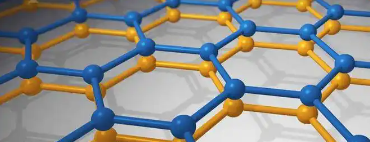
However, conventional imaging materials and sensors face inherent limitations: signal-to-noise ratio, sensitivity, and speed of response. This is where graphene, with its exceptional electrical, optical, and mechanical properties, emerges as a game-changer for next-generation medical imaging.
Why Graphene? Key Properties Relevant to Imaging
-
Extraordinary Electrical Conductivity
-
Graphene offers ultra-fast charge mobility, enabling rapid signal transfer in imaging detectors and sensors.
-
-
High Optical Transparency and Tunability
-
Nearly 97% transparent, graphene can be integrated into optical imaging windows and photodetectors without blocking light.
-
-
Flexibility and Thinness
-
A single-atom-thick graphene layer can be seamlessly incorporated into wearable and implantable imaging sensors.
-
-
Biocompatibility
-
Functionalized graphene materials can be engineered to interact safely with biological systems, useful in in-vivo imaging applications.
-
-
Enhanced Signal-to-Noise Ratio
-
Due to its low intrinsic noise and high sensitivity, graphene-based detectors can achieve superior resolution at lower radiation doses.
-
Applications of Graphene in Medical Imaging
1. Graphene-Enhanced MRI Contrast Agents
-
Graphene oxide and functionalized graphene nanosheets can be used as contrast-enhancing carriers in MRI.
-
These nanomaterials improve imaging clarity of tumors, brain tissue, and vascular structures.
-
Studies show graphene-based contrast agents can outperform traditional gadolinium-based agents in sensitivity.
2. Graphene in Ultrasensitive PET Detectors
-
PET requires highly sensitive photon detectors. Graphene-based photodetectors provide:
-
Faster response times
-
Lower dark noise
-
Improved time-of-flight (TOF) resolution
-
-
This leads to clearer images of metabolic activity, crucial for cancer diagnostics.
3. Graphene for Optical and Infrared Imaging
-
Graphene’s broadband light absorption makes it suitable for infrared and near-infrared (NIR) imaging devices.
-
Applications include:
-
Real-time vascular imaging during surgery
-
Retinal imaging for early detection of eye diseases
-
4. Graphene in Wearable Imaging Systems
-
Flexible graphene films can be integrated into skin-mounted ultrasound patches or portable diagnostic scanners.
-
Enables bedside imaging, continuous monitoring, and point-of-care diagnostics.
5. X-ray and CT Detector Improvements
-
Graphene’s high electron mobility improves X-ray sensor response times.
-
Potential to reduce radiation doses for patients while maintaining sharp resolution.
6. Functionalized Graphene for Molecular Imaging
-
By attaching biomolecules or nanoparticles to graphene, it can act as a targeted imaging probe.
-
Useful in fluorescence imaging or photoacoustic imaging to detect cancerous tissues at molecular levels.
Case Studies and Research Advances
-
Graphene Photodetectors for CT Imaging
-
Lab-scale prototypes using graphene sensors achieved 10x faster imaging speeds compared to silicon detectors.
-
-
Graphene Contrast Agents in Brain Imaging
-
Graphene-based nanosheets provided clearer MRI scans of brain tumors, with improved safety profiles over gadolinium.
-
-
Flexible Ultrasound Patches
-
Research groups are exploring graphene-polymer composites for wearable ultrasound patches that can image organs and muscles continuously without bulky equipment.
-
Advantages Over Conventional Materials
| Feature | Traditional Imaging Materials | Graphene-Based Materials |
|---|---|---|
| Electrical Conductivity | Moderate (Si, metals) | Ultra-high |
| Transparency | Limited (blocking in IR/NIR) | Up to 97% transparent |
| Sensitivity | High noise floor | High sensitivity, low noise |
| Flexibility | Rigid, brittle | Flexible, ultra-thin |
| Radiation Dose Requirement | High (CT, X-ray) | Lower possible with graphene detectors |
| Biocompatibility | Limited, risk of toxicity | Tunable with functionalization |
Challenges and Considerations
-
Scalability of Fabrication
-
High-quality, defect-free graphene production at scale is still a bottleneck.
-
-
Biocompatibility and Safety
-
Long-term studies are needed to ensure graphene-based contrast agents are safe for human use.
-
-
Integration with Existing Systems
-
Graphene detectors must be engineered to fit within current MRI, PET, and CT hardware without disrupting workflows.
-
-
Cost Efficiency
-
Transitioning from lab prototypes to mass-market clinical devices requires significant cost reductions.
-
Future Outlook
-
Next-Gen MRI and PET: With graphene-based contrast agents and detectors, medical imaging could achieve unprecedented resolution and sensitivity.
-
Wearable Imaging Devices: Portable, graphene-based systems may revolutionize telemedicine and remote diagnostics.
-
AI + Graphene Imaging: Combining graphene-enabled high-resolution imaging with AI-driven analysis could transform early disease detection.
-
Hybrid Materials: Graphene combined with CNTs or quantum dots may open new pathways in multi-modal imaging systems.
By 2030, graphene-enabled imaging devices could become standard in hospitals, delivering faster, safer, and more accurate diagnostics across oncology, neurology, and cardiology.
Graphene is redefining the possibilities of medical imaging by offering higher sensitivity, faster response, and improved safety compared to conventional materials. From MRI contrast agents to PET detectors and wearable ultrasound devices, graphene holds the potential to transform how diseases are detected and treated.
Although challenges remain in scalability, integration, and regulatory approval, rapid progress in graphene research suggests that its role in next-generation medical imaging will be profound.

